
Strange new worlds
Art from the science of bioengineering


Core to the research of the Auckland Bioengineering Institute, at Waipapa Taumata Rau, University of Auckland, is the application of engineering expertise to the complex structures and systems in human and animal biology and physiology. Researchers use a range of imaging techniques to gain insight into how these structures and systems work.
The goal is then to create digital twins for crucial aspects of human physiology. The ambition is for the digital twins to take away the guesswork and reduce the risk of surgery and clinical therapies. As part of this work researchers have created novel ways to visualise human and animal physiology.
Justin Fenandez on Muscles and bones:
"This image explores the interface of muscles and bones at the microscopic level. We often know about muscles connecting to bones but rarely ever see this complex connection. At the microscopic scale it looks like a galaxy that we are exploring.
"The blue region at the bottom is bone showing an osteon, a functional unit of bone. The black hole is a Haversian canal and the little dots around the rings are osteocytes that are mechanosensitive receptors that sense bone strain and initiate bone remodelling and adaptation to occur. The brown material across the top is the muscle tissue interfacing with the bone.
"Our group was interested in how pelvic bone adapts to the forces of pelvic muscles and everyday loads. These pelvic loads shape the architecture of bone and helps our bones cope with their mechanical environment.
“The image reminds me that there is a whole universe at the microscopic level of bones and muscles that we are still exploring."
This 3D microCT image shows a rare moment when a rat embryo is held within the amniotic sac, hanging from the placenta, resembling a blossom beginning its life. To model the development of the heart before birth, researchers carefully harvest rat embryos at the early days of gestation. Imaging techniques capture the structural and functional dynamics of the heart tissue at different biological levels from cell to whole organism ensuring the embryo remains intact.

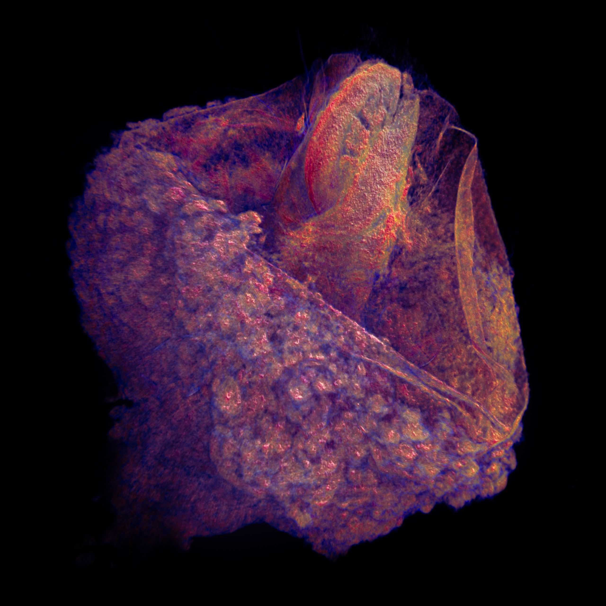


Gregory Sands says, " An uninterrupted supply of oxygenated blood is required for optimum functionality of the heart - this is provided by coronary circulation. The feathery nature is because those lines are each an individual vessel. It seems to swirl because the capillary directions change across the heart wall, which is because the muscle fibres have a rotation in their orientation across the heart wall.
This rotation of fibre orientation is one of the main reasons that the heart is so efficient at pumping blood. Without this rotation, the heart would not be able to pump blood at all.
These images are some of the most detailed images of cardiac microvasculature ever acquired, with the challenge being to maintain enough resolution to see individual vessels (1-2 microns)."

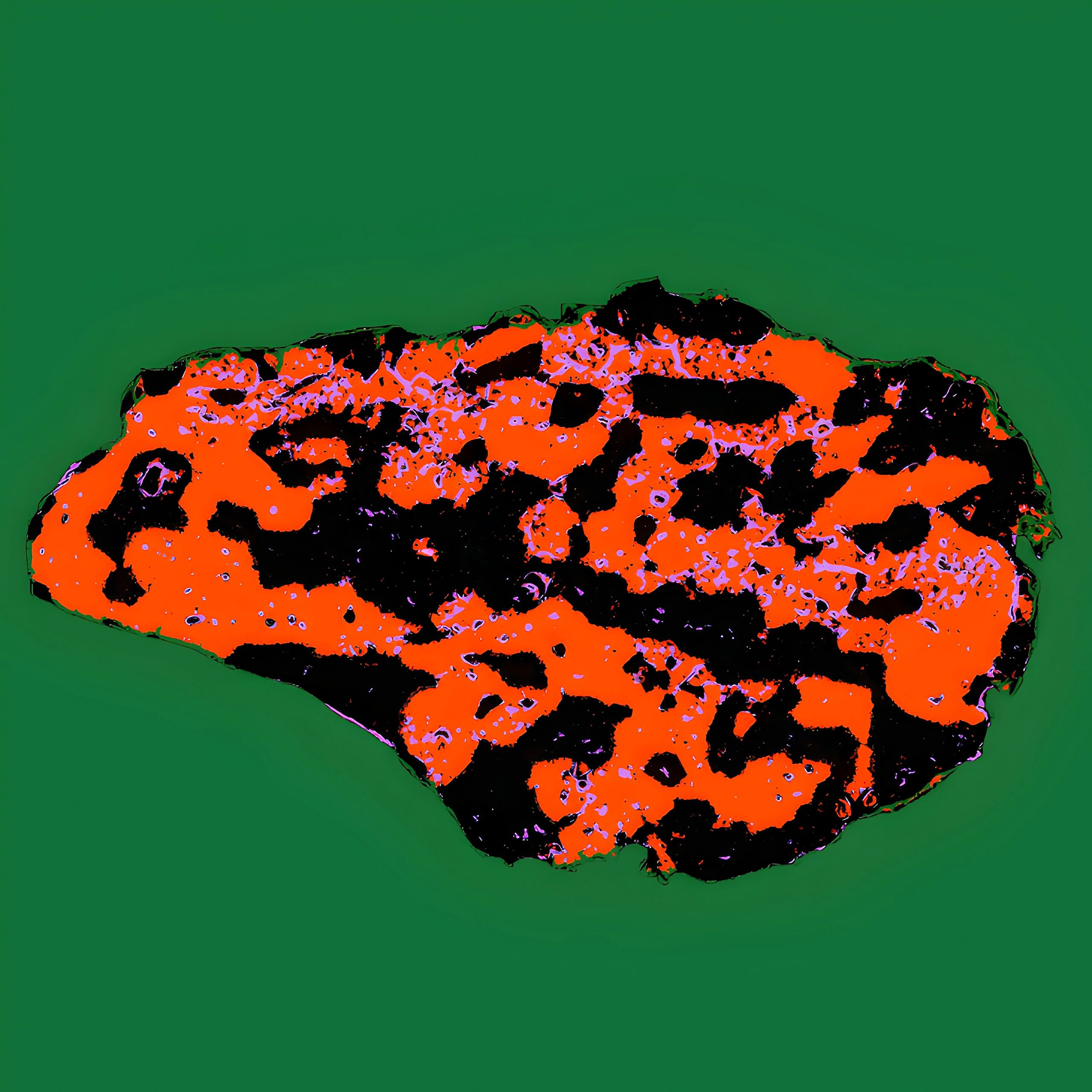
Your heart tirelessly pumps over 10,000 litres of blood through your body daily. This incredible pump is powered by countless heart cells. This image depicts a human heart cell as a vibrant, three-colour segmented transmission electron microscopy image, highlighting the distinct regions of mitochondria, myofibrils, and other cellular components.
Shashika Chamod says," Myofibrils are red, mitochondria and mitochondrial clusters in black, and other structures in purple. I used the colour to quantify features in diabetic and non-diabetic heart patients."

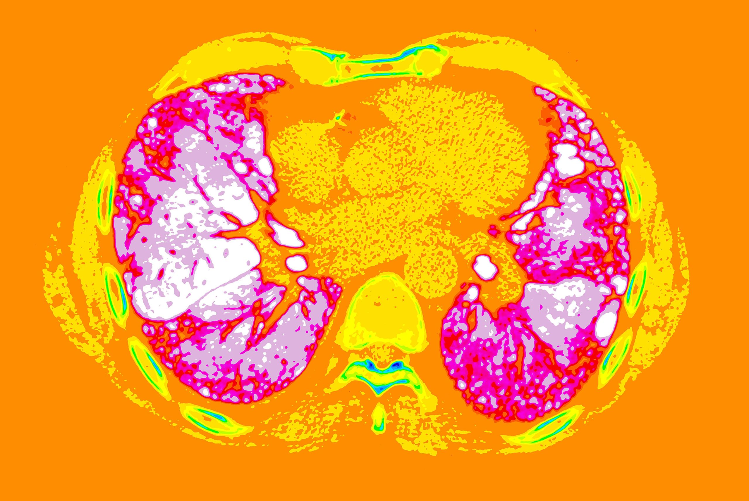
Joyce John says: "This precious pair of organs, vacuum-packed within the thoracic cavity, are responsible for oxygenating blood and removing carbon dioxide for the whole body.
The volume of the tissues contained within is only about 3 litres but the surface area is enough to cover a tennis court. The lung tissues can be scarred or remodelled by various diseases. The scarred tissues can be seen using computed tomography. They form ‘artistic patterns’ which can be distinguished from the normal tissue.
Here, the sugar-candy-coloured tissues are scarred due to idiopathic pulmonary fibrosis - the brighter the pink, the more scarring."
Associate Professor Peng Du says,"Sensors make contact with the body to record biological signals. Conventionally, sensor material is made of metal such as titanium, but through a chemical process called ‘doping’, non-conducting ‘plastic’ polymers can be made to carry electric current.
This was the legacy bequeathed to us by the late New Zealand Nobel laureate Professor Alan MacDiarmid. Conducting polymer is now an exciting new frontier of material chemistry, with many applications for improved signal acquisition and biological capability.
Conventional sensors detect one type of signal at a time, but these sensors can be 'tuned' to detect different types of signals, which means within a single patch we may be able to detect multiple types of signals as once. This reduces the size of the device, whether it is implanted or applied outside the body."


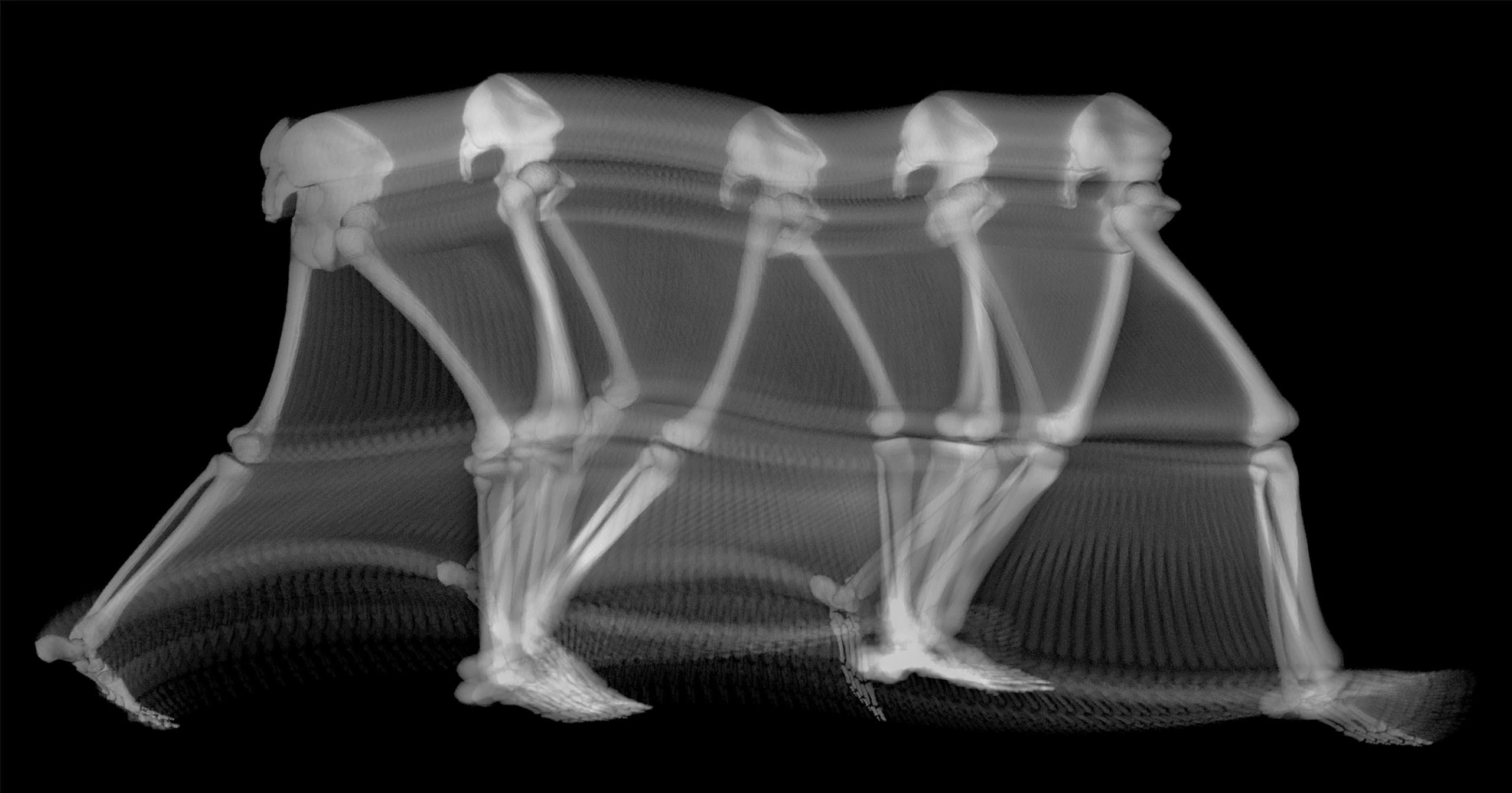
Duncan Bakke says: "When we conduct research (like modelling human gait) the end result may feel anticlimactic. Fake legs taking fake steps. But each step is the culmination of thousands of hours of work from dozens of researchers.
"Each of the bones visible in this image were generated using a shape modelling workflow carefully crafted using models of hundreds of people.
"The steps were recorded after training from several people, themselves trained for years - simulated in an open-source engine that’s taken a decade to develop. A giant leap, composed of a million tiny motes of progress. All for this one small step."

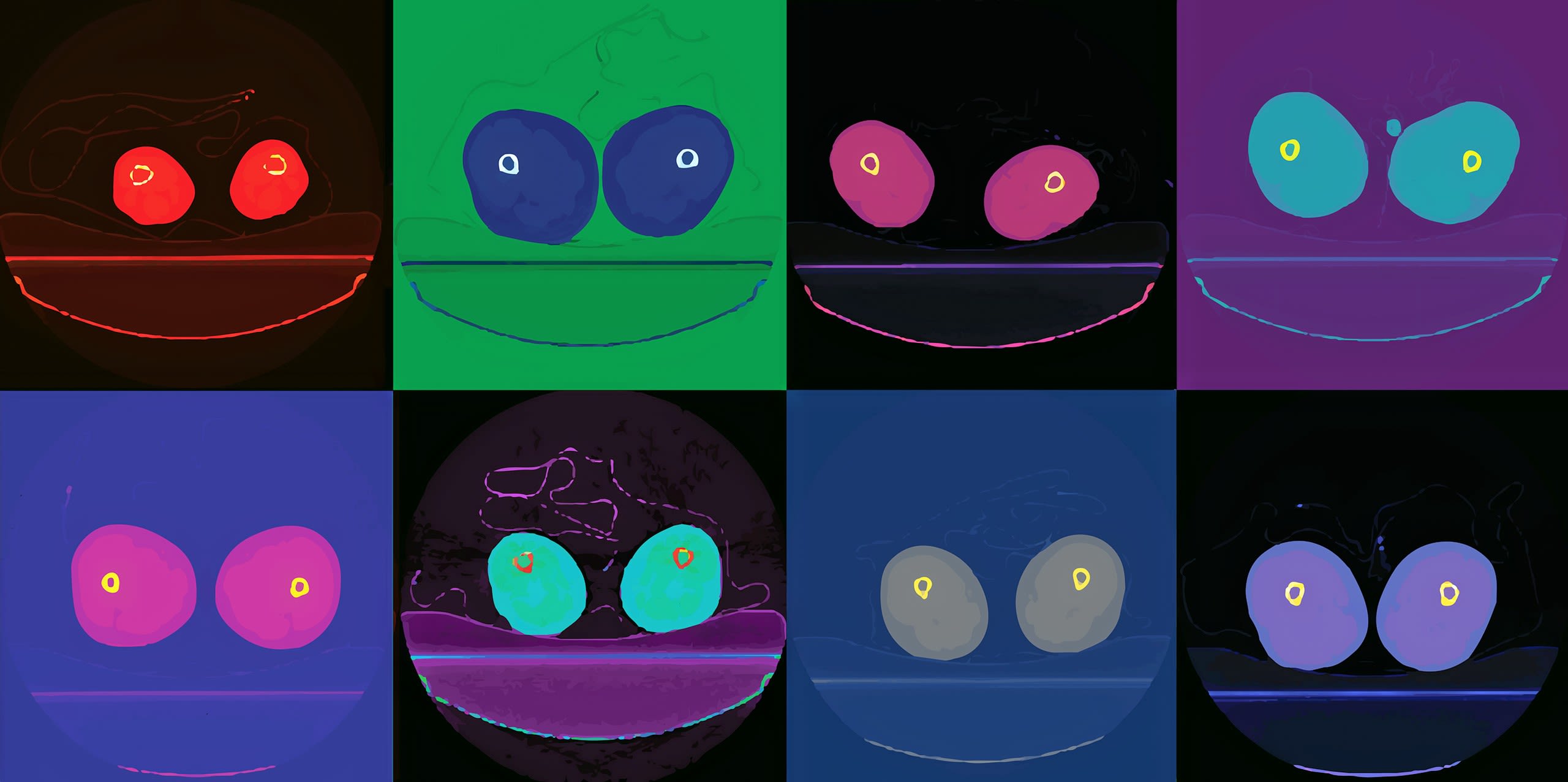
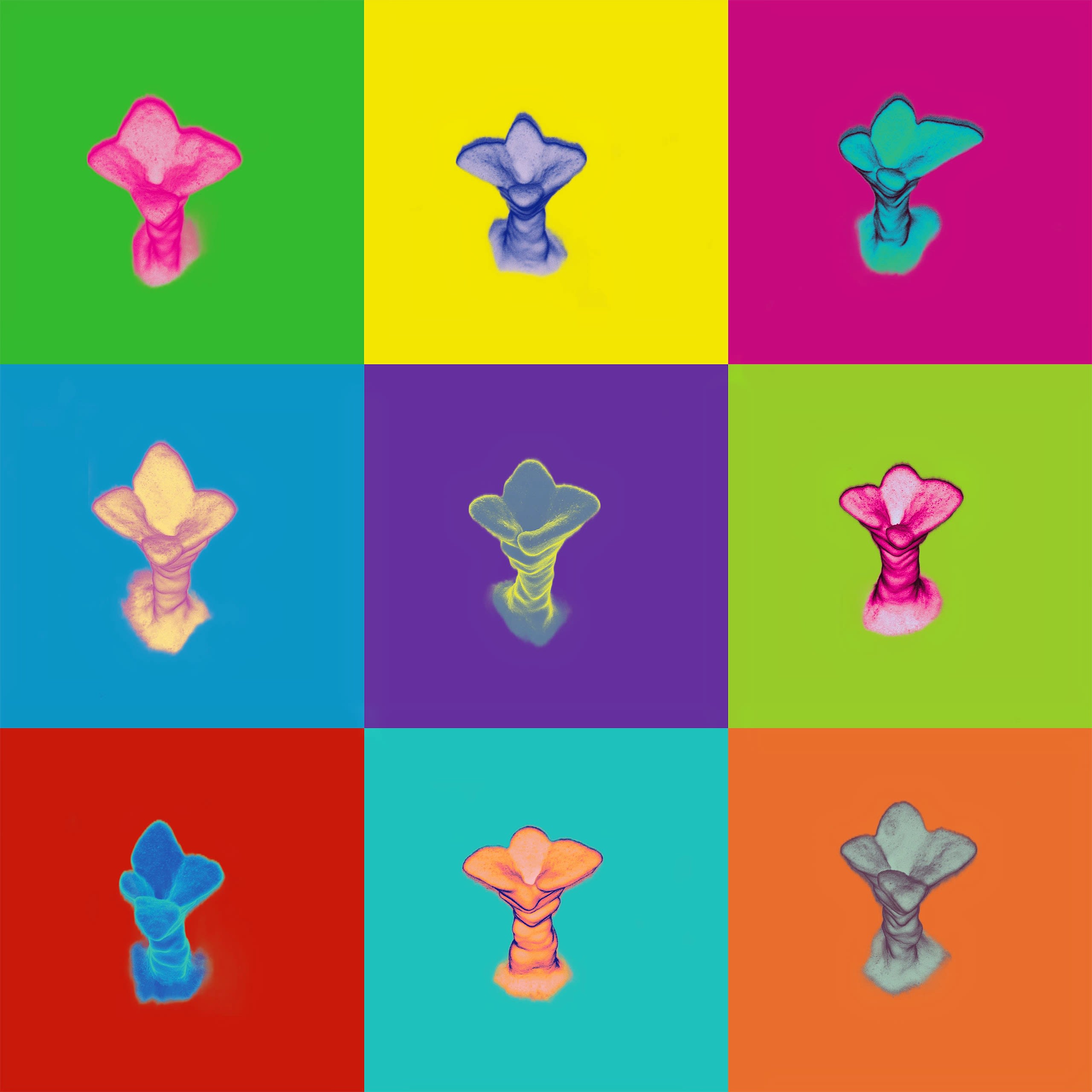
The team explains: "We started with a dataset of computer tomography images of human legs. The curve of the exam table under the legs looked like a smile, and suddenly the femurs looked like eyes.
All of a sudden we were looking upon a dataset of cute googly eyed faces. We then used color maps from the Matplotlib Python library to create an image inspired by the Marilyn Monroe series by Andy Warhol."

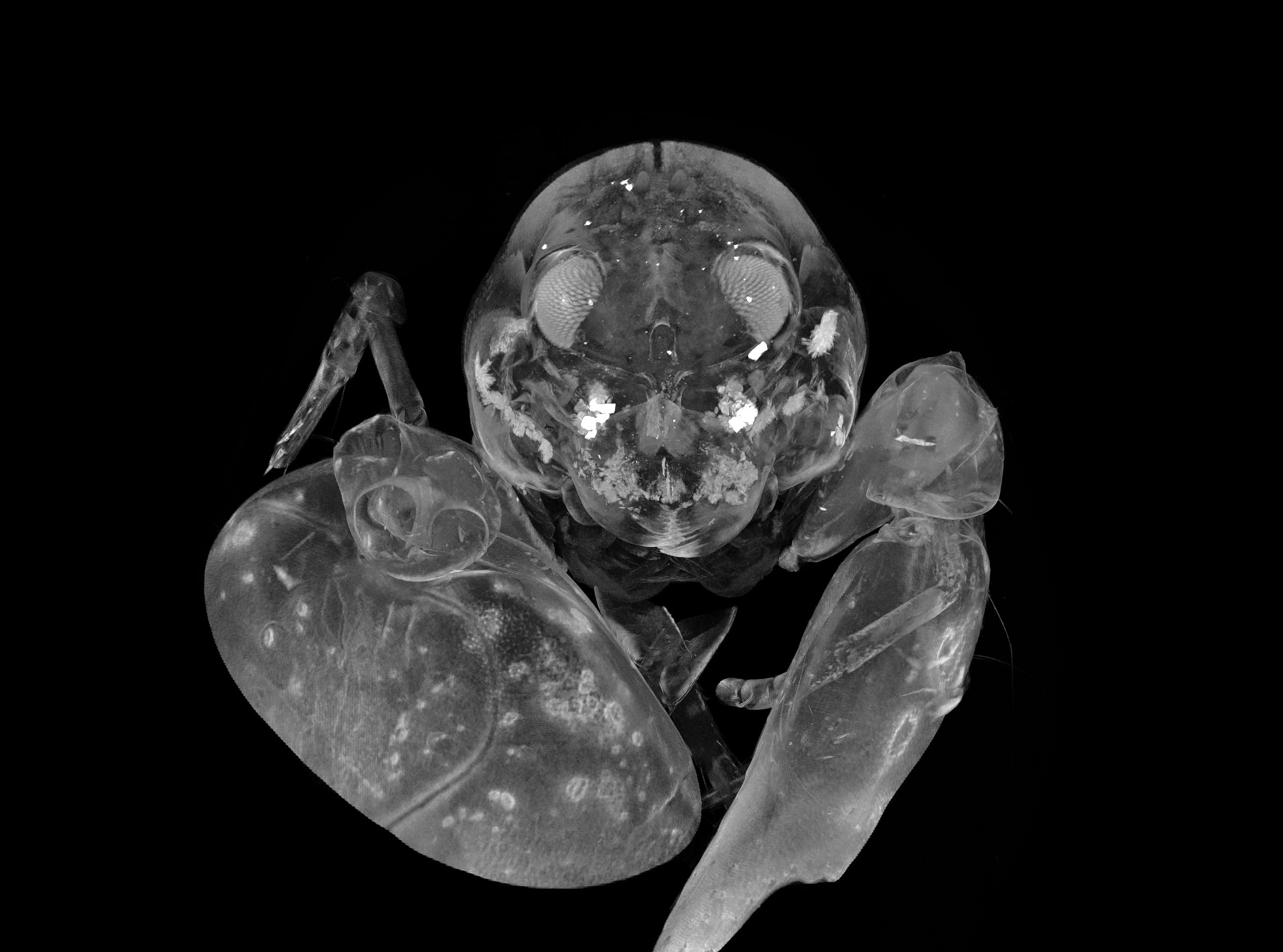
Snapping shrimps are social creatures. One of their first legs bears a voluminous claw. The snap of the claw creates a cavitation bubble so loud that it is detectable for hundreds of meters underwater. The MicroCT image reveals the structure and features of the shrimp's compound eyes and the bright statocyst complex, how shrimps might 'hear' acoustic stimuli underwater.

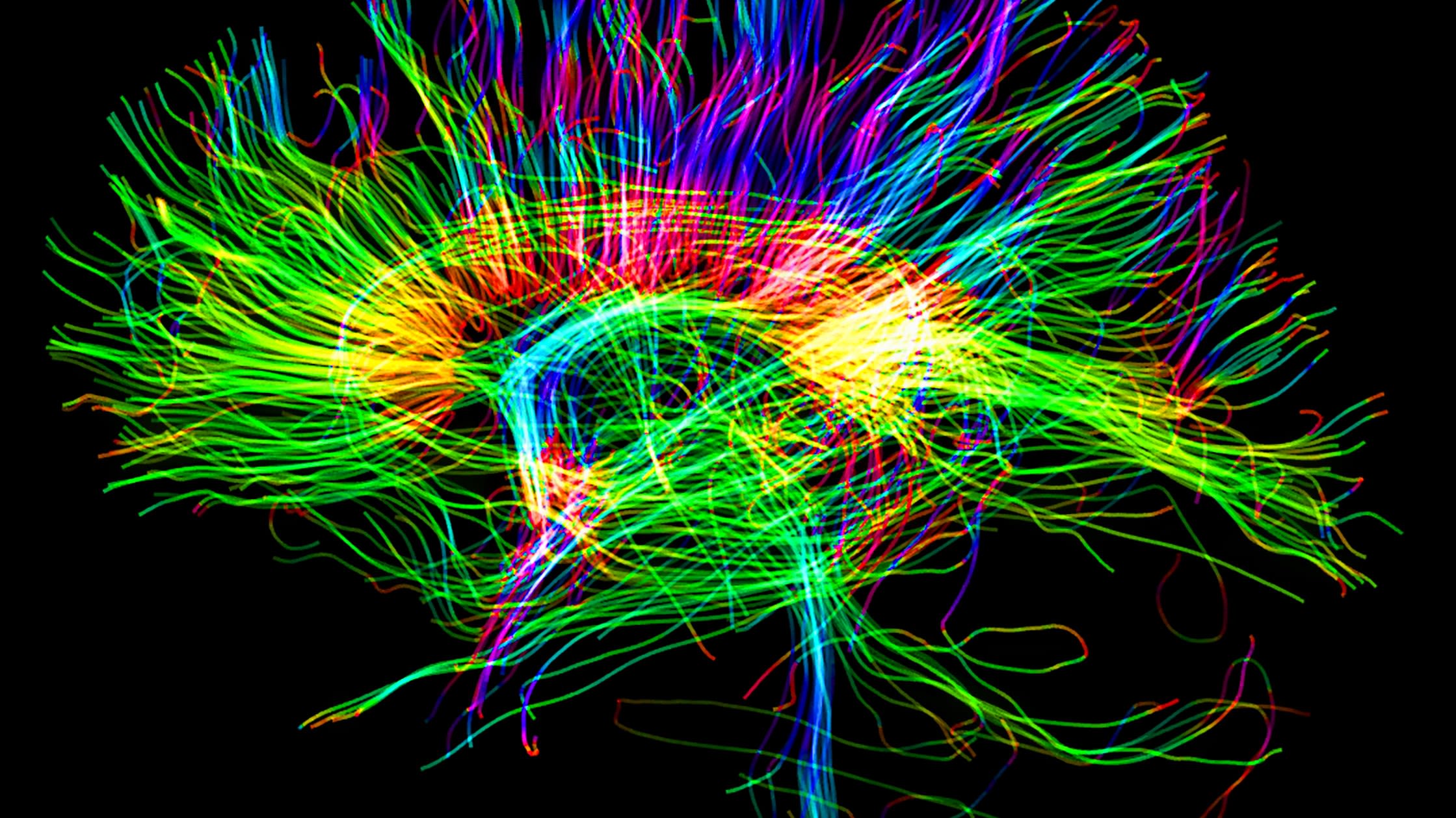
Maryam Tayebi says, "Billions of neural fibers in your brain act as a relay to coordinate communication between different regions. Thanks to an advanced medical imaging technique - tractography - these fiber tracts can be visualised in a unique fashion. Just by slightly changing your view, you can find the extraordinary patterns that these fibers are making in your brain."